vivoScreen
Automated High-Throughput, High-Content Imaging System for C. elegans Researchers

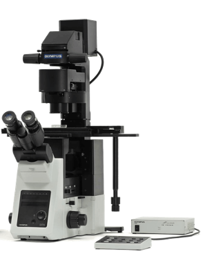
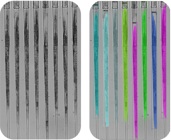
vivoScreen is a complete imaging solution enabling high-resolution imaging of C. elegans at high-throughput scales. At the heart of the system is the vivoChip-24x, a multiwell plate format microfluidic device that can immobilize and laterally align up to 40 worms from 24 different populations for high-resolution imaging, for a total of nearly 1,000 animals in a single imaging session. When combined with the fully motorized microscope and intuitive experiment design and image acquisition, the vivoScreen opens up new experimental avenues not possible with traditional microscopes or imaging systems.
High-Content C. elegans Imaging at Plate Reader Speeds
High-Content C. elegans Imaging at Plate Reader Speeds
| Image whole bodies of ~1000 worms from 24 populations at ~0.4 µm pixel resolution in 10 minutes using vivoImager and vivoChip-24x* |
| Image a 384-well plate in 10 minutes (1 image per well) using vivoImager |
| Automatically calculate ~1000 worm lengths/volumes in 1 minute with vivoAI |
| Automatically calculate ~1000 worm aggregate counts and sizes in 2 hours with vivoAnalyzer |

A Future-Proof System Built to Help Now
A Future-Proof System Built to Help Now
While it is excellent for general research use, the standard vivoScreen build has been specialized for imaging using the microfluidic vivoChip-24x, making vivoScreen the only system capable of high-throughput and high-resolution of imaging of C. elegans. Large field-of-view optics, a state-of-the-art camera, motorized components, and a modular light path make the vivoScreen imaging system both powerful enough to fulfill a wide range of daily research needs and flexible enough to evolve as research requirements change.

| Software-controllable 6-position objective mount, 8-position fluorescence cube deck, and 7-position long-working-distance condenser turret | |
| Readily accessible light path for easy changing of optical components as your research needs evolve |
| Water- and oil-immersion objective compatibility | |
| Fluorescence, brightfield, DIC, and phase contrast imaging compatibility | |
| Software-controllable LED light sources for instantaneous and stable illumination |
Software Tools for Automated Imaging and Analysis
Software Tools for Automated Imaging and Analysis
| Manual and automated imaging modes with autofocus and worm detection for use with vivoChip-24x | |
| Time-lapse, Z-stack, and tiling image capture options | |
| Expanded protocols for imaging well plates and other standard sample vessel formats with future software updates |
| Point-and-click-based user-defined phenotype and feature identification workflows for custom assay analysis | |
| Fully-automated AI-assisted analysis of supported phenotypes and features | |
| Analysis output data exportable to a spreadsheet format |


